Hidetaka Arimura Laboratory, Kyushu University
Research
In our AI-based oncology researches, we are making use of an eclipse radiation treatment planning system (Varian Medical Systems) for delineation of tumor contours, treatment planning, and so on.
Papers 2026-1-Cui

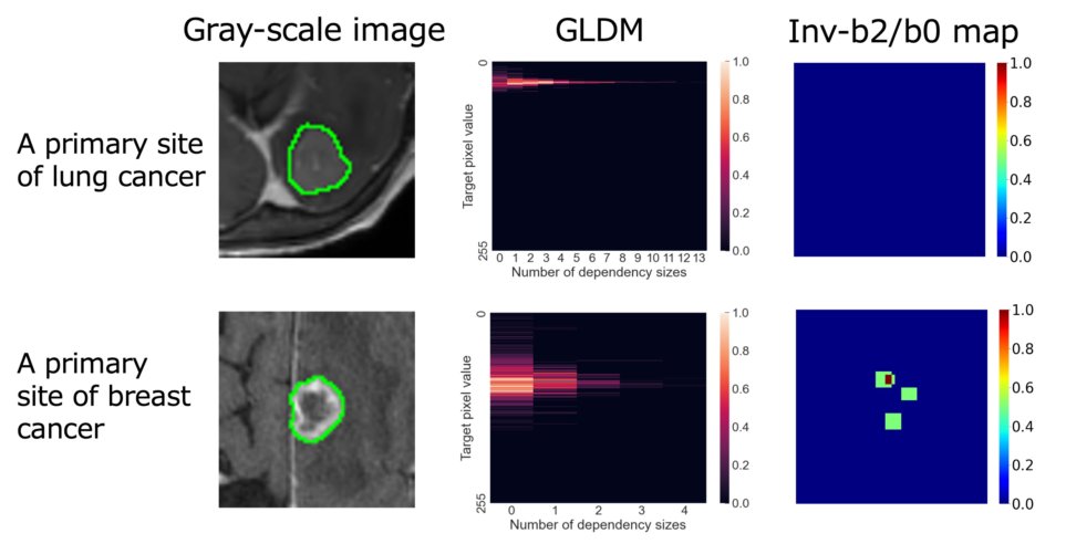
Interpretable classification model of lung nodules into three morphological
types based on consolidation-to-tumor-ratio-related features on computed
tomography images
Yunhao Cui, Hidetaka Arimura, Yuko Shirakawa, Tadamasa Yoshitake, Yu Jin, Yoshiyuki Shioyama, Hidetake Yabuuchi
Health and Technology (accepted on April 02, 2026)
Purpose:We explored automated classification models of lung nodules into
three morphological types [solid, part-solid, and pure ground-glass nodules
(pGGN)] based on computed tomography (CT) using features related to the
three-dimensional (3D) consolidation-to-tumor ratio (CTR), which is the
maximum diameter ratio of consolidation (=solid component) to a whole tumor.
Conclusions:This study suggests that the proposed interpretable classification
model using CTR-related features could robustly predict morphological nodule
types.
Keywords: Deep learning, consolidation-to-tumor-ratio-related features, lung cancer,
nodule types
Papers 2025-9-Anton
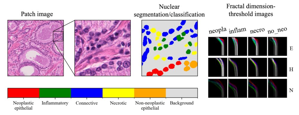
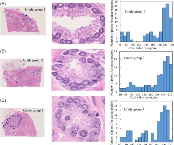
Fractal dimensions for tumour-related cell types of prostate cancer on
histopathology images using multiple-threshold box counting algorithm
Anton Schwarz, Hidetaka Arimura, Yunhao Cui, Shun Shimabukuro, Qijing Lin, Yu Jin, Satoshi Kobayashi, Takashi Matsumoto, Masaki Shiota, Masatoshi Eto, Yoshinao Oda
Biophysics and Physicobiology
Advance online Publication on October 18, 2025
DOI: https://doi.org/10.2142/biophysico.bppb-v22.0026
Histopathological images taken from prostate cancer biopsies are segmented
by five tumour-related cell types as shown in the top left three images.
The distribution of pixel values in the image exhibits self-similarity
and is described by the fractal dimension (FD). We found that FD-threshold
images like the right images can be leveraged to predict low and high grades
of prostate cancer. Here, E: Eosin, H: Hematoxylin, N: Normalised image,
neopla: Neoplastic epithelial, inflam: Inflammatory, necro: Necrotic, and
no_neo: Non-neoplastic epithelial.
Keywords: Fractal dimension, prostate cancer malignancy, histopathology image,
multiple-threshold box counting
Papers 2025-8-Yagiz
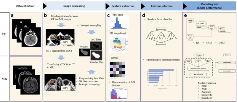
Non-invasive Prediction of Secondary Enucleation Risk in Uveal Melanoma
Based on Pretreatment CT and MR Imaging Prior to Stereotactic Radiotherapy
Yagiz Yedekci, Hidetaka Arimura, Yu Jin, Melek Tugce Yilmaz, Takumi Kodama,
Gokhan Ozyigit, Gozde Yazici.
Strahlentherapie und Onkologie
Published: 08 August 2025
https://doi.org/10.1007/s00066-025-02449-1
*Green Open Access*
Purpose: The aim of this study was to develop a radiomic model to non-invasively
predict the risk of secondary enucleation (SE) in patients with uveal melanoma
(UM) prior to stereotactic radiotherapy using pretreatment computed tomography
(CT) and magnetic resonance (MR) images.
Keywords: Machine learning in medicine, Predictive model, Prognosis prediction, Radiomics,
Side effect prediction
Papers 2025-7-Mitsushima
Time-variant tumor growth trajectory models for in silico randomized controlled
trials for patients with early-stage non-small cell lung cancer in optimizing
stereotactic body radiation therapy
Kazuki Mitsushima,Hidetaka Arimura, Yuko Shirakawa, Takumi Kodama, Tadamasa
Yoshitake.
Health and Technology
Published 05 August 2025
*Geen open access*
https://doi.org/10.1007/s12553-025-01005-2
Purpose: Applying new treatments to real patients to verify therapeutic
efficacy may induce various risks, such as critical adverse events. Additionally,
there are ethical and financial issues in real-world randomized controlled
trials (RCTs). This study aimed to develop mathematical models of time-variant
tumor growth trajectories (TGTs) for in silico RCTs targeting patients
with stage I non-small cell lung cancer (NSCLC) to optimize stereotactic
body radiation therapy (SBRT).
Keywords: Lung cancer, Tumor growth trajectory, In silico simulations, Radiation
therapy, Differential equations
Papers 2025-6-Kamezawa
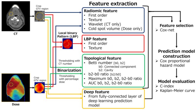
Radiodosiomics Prediction of Treatment Failures Prior to Chemoradiotherapy
in Head-and-Neck Squamous Cell Carcinoma
Hidemi Kamezawa, Arimura Hidetaka.
Applied Sciences, 2025, 15(12), 6941
Published: 19 June 2025
https://doi.org/10.3390/app15126941
Abstract: Predicting treatment failure (TF) in head-and-neck squamous cell
carcinoma (HNSCC) patients before treatment can help in selecting a more
appropriate treatment approach. We investigated a novel radiodosiomics
approach to predict TF prior to chemoradiation in HNSCC patients.
Keywords: Treatment failure prediction, Head-and-neck squamous cell carcinoma,
Topology, Radiodosiomics
Papers 2025-5-Jin Y

Multiscale-fusion models with genomic, topological and pathomic features to predict response to radiation therapy for non-small cell lung cancer patients
Yu Jin, Hidetaka Arimura, Takeshi Iwasaki, Takumi Kodama, Noriaki Yamamoto, Yunhao Cui, Yoshinao Oda.
Laboratory Investigation, Published online, 12 June 2025
Laboratory Investigation Vol. 105, Issue 10104204, October 2025
https://doi.org/10.1016/j.labinv.2025.104204
Abstract: We investigated fusion models with multiscale features in histopathology
images to predict response to radiation therapy for patients (responders)
with non-small cell lung cancer.
Keywords: Fusion model, Histopathology image, Lung cancer, Radiation therapy, Topology
Papers 2025-4-Shibayama

Can Online-Adaptive Radiation Therapy Eliminate Intra-Fractional Deformation
in Gastric Mucosa-Associated Lymphoid Tissue Lymphoma?
Yusuke Shibayama, Hidetaka Arimura, Taka-aki Hirose, Masanori Takaki, Jun-ichi
Fukunaga, Tadamasa Yoshitake, Toyoyuki Kato, Kousei Ishigami.
Practical Radiation Oncology 2025
Articles in Press: June 09, 2025
https://doi.org/10.1016/j.prro.2025.05.008
Purpose: We hypothesized that online adaptive radiation therapy (oART) could eliminate errors associated with interfractional deformation in gastric mucosa-associated lymphoid tissue (MALT) lymphoma, but errors in intrafractional deformation remained in 6 directions (anterior, posterior, superior, inferior, left, and right). This study aimed to quantify the anisotropic deformation errors of the clinical target volume (CTV) for MALT lymphoma using oART to determine deformations in the planning target volume (PTV) margins.
Keywords: Online Adaptive Radiation Therapy; Interfractional Deformation; Intrafractional
Deformation; Gastric Mucosa-Associated Lymphoid Tissue Lymphoma
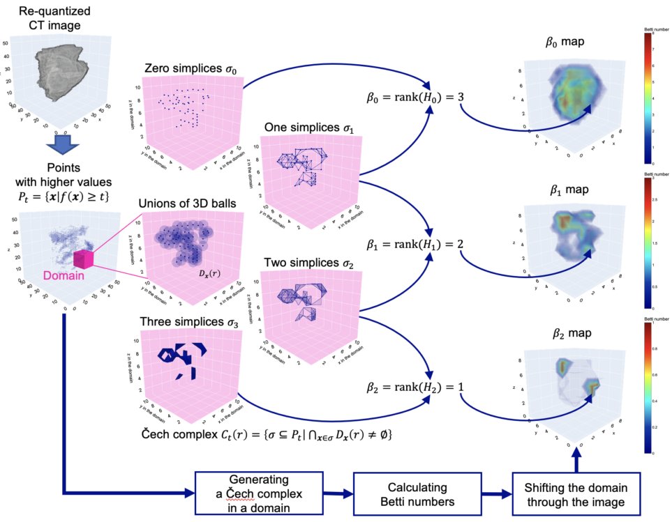
Papers 2025-3-Iwasaki

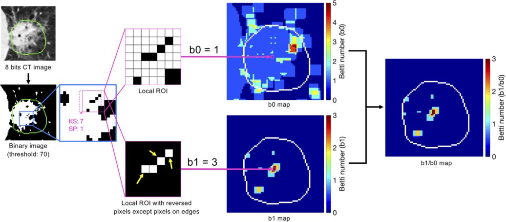
Predictive models of severe disease in patients with COVID-19 pneumonia
at an early stage on CT images using topological properties
Takahiro Iwasaki, Hidetaka Arimura, Shohei Inui, Takumi Kodama, Yun Hao
Cui, Kenta Ninomiya, Hideyuki Iwanaga, Toshihiro Hayashi, Osamu Abe.
Radiological Physics and Technology 2025 Vol.18, 534-546.
Published: 28 April 2025
https://doi.org/10.1007/s12194-025-00906-1
Abstract: We aimed to construct predictive models of SVD in patients with
COVID-19 pneumonia at an early stage on computed tomography (CT) images
using SVD-specific features that can be visualized on accumulated Betti
number (BN) maps. BN maps (b0 and b1 maps) were generated by calculating
the BNs within a shifting kernel in a manner similar to a convolution
Keywords: COVID-19, Severity, Topological features, Accumulated Betti number map, Predictive model
Papers 2025-2-Shibayama

Explainable Radiomics based on Association of Histopathological Cell Density
and Multiparametric MR Radiomic Features for High-Risk Stratification of
Prostate Cancer Patients
Yusuke Shibayama, Hidetaka Arimura, Yukihisa Takayama, Fumio Kinoshita, Dai Takamatsu, Akihiro Nishie, Satoshi Kobayashi, Takashi Matsumoto, Masaki Shiota, Masatoshi Eto, Yoshinao Oda, Kousei Ishigami.
Magnetic Resonance Materials in Physics, Biology and Medicine
Published: 24 April 2025
https://doi.org/10.1007/s10334-025-01250-6
*Green Open Access*
Objective: This study aimed to develop an explainable radiomics model for
stratifying prostate cancer (PCa) patients with high-risk disease via investigation
of the association between cell density (CD) in the PCa region on histopathological
images and multiparametric MR (mpMR) radiomics features.
Keywords: Explainable radiomics, Prostate cancer, Cell density, Histopathological
images, Multiparametric MR images
Papers 2025-1-Kodama

Topological radiogenomics based on persistent lifetime images for identification
of epidermal growth factor receptor mutation in patients with non-small
cell lung tumors
Takumi Kodama; Hidetaka Arimura; Tomoki Tokuda; Kentaro Tanaka; Hidetake
Yabuuchi; Nadia Fareeda Muhammad Gowdh; Chong Kin Liam; Chee Shee Chai;
Kwan Hoong Ng.
Computers in Biology and Medicine Volume 185, February 2025, 109519
https://doi.org/10.1016/j.compbiomed.2024.109519
Highlights
*Persistent lifetime (PLT) images have been newly proposed to characterize
the spatial heterogeneity of risk factors for epidermal growth factor receptor
(EGFR) mutation in patients with non-small cell lung cancer (NSCLC).
*PLT images explicitly enhanced the locations and persistent contrasts of topological components (connected and hole components) corresponding to EGFR mutant traits.
*2D-PLT features can be radiogenomic imaging biomarkers to show robust
and high identification of EGFR mutation-positive patients compared with
conventional features.
Keywords: Radiogenomics, Persistent lifetime image EGFR mutation, Precision medicine
Papers 2024-2-Hamasaki

Noninvasive machine-learning models for the detection of lesion-specific
ischemia in patients with stable angina with intermediate stenosis severity
on coronary CT angiography
Hiroshi Hamasaki, Hidetaka Arimura, Yuzo Yamasaki, Takayuki Yamamoto, Mitsuhiro
Fukata, Tetsuya Matoba, Toyoyuki Kato, Kousei Ishigami.
Physical and Engineering Sciences in Medicine
(Accepted: 05 Dec.2024, published online ahead of print on December 30,
2024)
https://doi.org/10.1007/s13246-024-01503-z
*Green Open Access*
Abstract:This study proposed noninvasive machine-learning models for the detection
of lesion-specific ischemia (LSI) in patients with stable angina with intermediate
stenosis severity based on coronary computed tomography (CT) angiography.
These findings suggest that LSI detection models with features extracted
from coronary CT angiography (CCTA) can noninvasively detect LSI in patients
with stable angina with intermediate stenosis severity.
Keywords: Noninvasive machine-learning, Lesion-specific ischemia, Coronary CT angiography
Papers 2024-1-Tong

Automated prediction of consolidation tumor ratio for stage I non-small
cell lung cancer from treatment planning CT images based on deep learning
segmentation models
YiZhi Tong, Hidetaka Arimura, Tadamasa Yoshitake, Yunhao Cui,Takumi Kodama,
Yoshiyuki Shioyama, Ronnie Wirestam, Hidetake Yabuuchi
Applied Sciences 2024 2024, 14(8), 3275. Published: 13 April 2024
https://doi.org/10.3390/app14083275
Abstract:This study aimed to propose an automated prediction approach of the consolidation
tumor ratios (CTRs) of part-solid tumors of patients treated with radiotherapy
on treatment planning Abstract:Tcomputed tomography images using deep learning
segmentation models. The findings suggest that the automated prediction
approach could be robust in calculating CTRs of part-solid tumors.
Keywords: Consolidation tumor ratio, Deep learning, Part-solid tumors, Independent
test, Non-small cell lung cancer (NSCLC)
Papers 2023-5-Egashira
Magnetic Resonance-Based Imaging Biopsy with Signatures Including Topological Betti Number Features for Prediction of Primary Brain Metastatic Sites
Mai Egashira, Hidetada Arimura, Kazuma Kobayashi, Kazutoshi Moriyama, Takumi
Kodama, Tomoki Tokuda, Kenta Ninomiya, Hiroyuki Okamoto, Hiroshi Igaki
Physical and Engineering Sciences in Medicine (Published: 21 August 2023)
https://doi.org/10.1007/s13246-023-01308-6
Abstract:This study incorporated topology Betti number (BN) features into the prediction
of primary sites of brain metastases and the construction of magnetic resonance
(MR)-based imaging biopsy (MRB) models. The significant features of the
MRB model were selected from those obtained from gray-scale and three-dimensional
wavelet-filtered images, BN and inverted BN (iBN) maps, and clinical variables
(age and gender). The results suggest that the BN signature boosted the
performance of MRB for the identification of primary sites of brain metastases
including small tumors.We investigated an approach for predicting recurrence
after radiation therapy using local binary pattern (LBP)-bas
Keywords: Imaging Biopsy, Betti number,Topology
Papers 2023-4-Cui
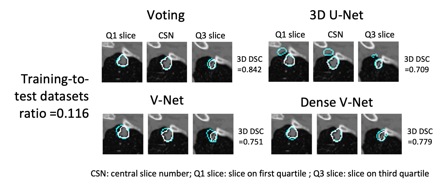
Deep learning model fusion improves lung tumor segmentation accuracy across variable training-to-test dataset ratios
Yunhao Cui, Hidetaka Arimura, Tadamasa Yoshitake, Yoshiyuki Shioyama, Hidetake Yabuuchi
Physical and Engineering Sciences in Medicine (Published: 07 August 2023)
https://doi.org/10.1007/s13246-023-01295-8
Abstract:This study aimed to investigate the robustness of a deep learning (DL)
fusion model for low training-to-test ratio (TTR) datasets in the segmentation
of gross tumor volumes (GTVs) in three-dimensional planning computed tomography
(CT) images for lung cancer stereotactic body radiotherapy (SBRT). Three
DL models, 3D U-Net, V-Net, and dense V-Net, were trained to segment the
GTV regions. Nine fusion models were constructed with logical AND, logical
OR, and voting of the two or three outputs of the three DL models. TTR
was defined as the ratio of the number of cases in a training dataset to
that in a test dataset. The voting fusion model achieved the highest DSCs
of 0.829 to 0.798 for all TTRs among the 12 models. The findings suggest
that the proposed voting fusion model is a robust approach for low TTR
datasets in segmenting GTVs in planning CT images of lung cancer SBRT.
Keywords: deep learning, lung cancer
Papers 2023-3-Jin
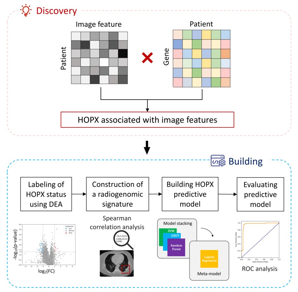
CT image-based biopsy to aid prediction of HOPX expression status and prognosis for non-small cell lung cancer patients
Yu Jin, Hidetaka Arimura, YunHao Cui, Takumi Kodama, Shinichi Mizuno, Satoshi Ansai
Cancers 2023, 15(8), 2220, Published: 10 April 2023
https://doi.org/10.3390/cancers15082220
Abstract: Recent studies have found that the HOPX gene functions as a tumor
suppressor, and its expression status influences patients’ survival in
NSCLC. This study established an imaging biopsy with the radiogenomic signatures
that links HOPX expression status and CT images to aid the prediction of
HOPX expression status and the prognosis for lung cancer patients. Detecting
gene expression status from CT images might be helpful to improve the accuracy
of wet biopsy.
Keywords:HOPX; CT image features; imaging biopsy; non-small cell lung cancer; radiogenomics
Papers 2023-2-Ninomiya
Three-dimensional topological radiogenomics of epidermal growth factor
receptor Del19 and L858R mutation subtypes on computed tomography images
of lung cancer patients
Kenta Ninomiya, Hidetaka Arimura, Kentaro Tanaka, Wai Yee Chan, Yutaro
Kabata, Shinichi Mizuno, Nadia Fareeda Muhammad Gowdh, Nur Adura Yaakup,
Chong-Kin Liam, Chee-Shee Chai, Kwan Hoong Ng
Computer Methods and Programs in Biomedicine (accepted on Apr 7, 2023)
https://doi.org/10.1016/j.cmpb.2023.107544
Abstract: The objective of this study was to elucidate a novel radiogenomics approach using three-dimensional (3D) topologically invariant Betti numbers (BNs) for topological characterization of epidermal growth factor receptor (EGFR) Del19 and L858R mutation subtypes. 3DBN features, which showed a radiogenomic association with the characteristics of the EGFR Del19/L858R mutation subtypes, yielded higher accuracy for subtype classifications in comparison with conventional features.
Keywords: radiogenomics, computational topology, molecularly targeted drugs, precision
medicine
Papers 2023-1-Ikushima
Topology-based radiomic features for prediction of parotid gland cancer
malignancy grade in magnetic resonance images
Kojiro Ikushima, Hidetaka Arimura, Ryuji Yasumatsu, Hidemi Kamezawa, Kenta
Ninomiya
Magnetic Resonance Materials in Physics, Biology and Medicine, (Published:
20 April 2023)
https://doi.org/10.1007/s10334-023-01084-0
Abstract:The malignancy grades of parotid gland cancer (PGC) have been assessed
for decision of treatment policies. Therefore, we have investigated the
feasibility of a topology-based radiomic features for prediction of parotid
gland cancer (PGC) malignancy grade in magnetic resonance (MR) images.This
study indicated that topology-based radiomic features could be feasible
for the noninvasive prediction of the malignancy grade of PGCs.
Keywords: Radiomic features, Topology, Parotid gland cancer, Malignancy grade
Papers 2022-6-Kamezawa
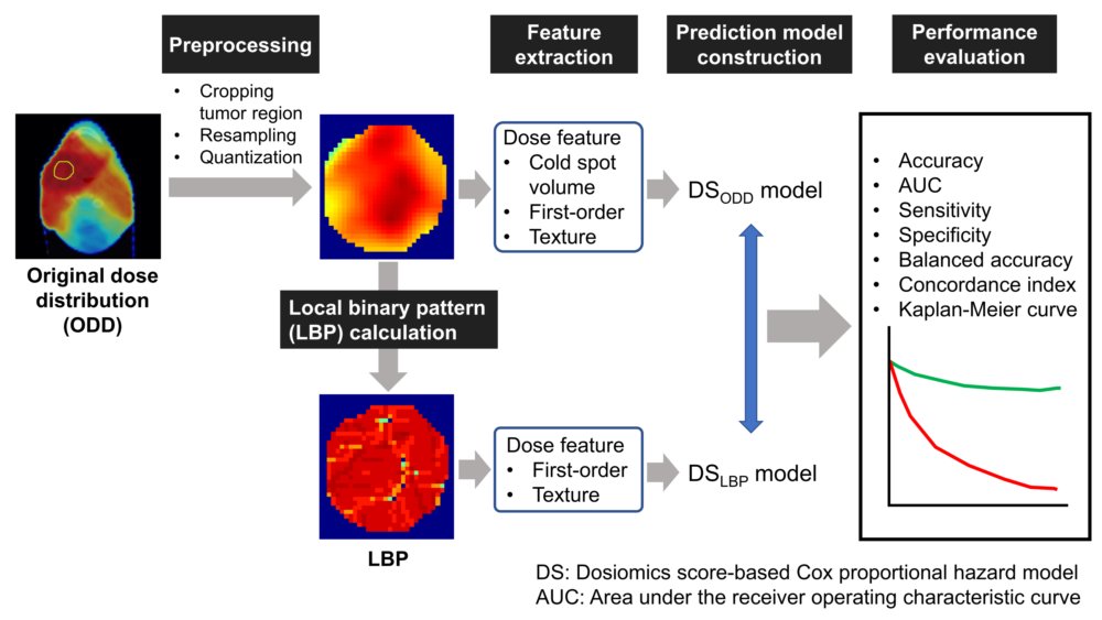
Recurrence prediction with local binary pattern-based dosiomics in patients
with head and neck squamous cell carcinoma
Kamezawa Hidemi, Arimura Hidetaka, et al.
Physical and Engineering Sciences in Medicine (Published: 05 December 2022)
https://doi.org/10.1007/s13246-022-01201-8
Abstract:We investigated an approach for predicting recurrence after radiation therapy using local binary pattern (LBP)-based dosiomics in patients with head and neck squamous cell carcinoma (HNSCC). LBP-based dosiomics models may be more accurate in predicting recurrence after radiation therapy in patients with HNSCC.
Keywords: recurrence prediction, head and neck carcinoma, local binary pattern, dosiomics
Papers 2022-5-Nagami
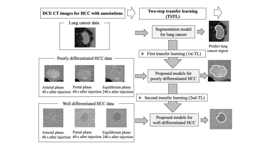
Dual segmentation models for poorly and well-differentiated hepatocellular
carcinoma using two-step transfer deep learning on dynamic contrast-enhanced
CT images
Noriyuki Nagami, Hidetaka Arimura, Junichi Nojiri, Cui Yunhao, Kenta Ninomiya,
Manabu Ogata, Mitsutoshi Oishi, Keiichi Ohira, Shigetoshi Kitamura, Hiroyuki
Irie.
Physical and Engineering Sciences in Medicine (Published: 05 December 2022)
https://doi.org/10.1007/s13246-022-01202-7
Abstract: The aim of this study was to develop dual segmentation models for poorly and well-differentiated hepatocellular carcinoma (HCC), using two-step transfer learning (TSTL) based on dynamic contrast-enhanced (DCE) computed tomography (CT) images. The proposed model using TSTL from the lung cancer dataset showed the potential to segment poorly and well-differentiated HCC regions on DCE-CT images.
Keywords: deep learning, hepatocellular carcinoma, dual segmentation, poorly
differentiated, well-differentiated, transfer learning
Papers 2022-4-Moriyama

Feasibility for prediction of primary cancer sites of brain metastases
based on Hessian index images
Kazutoshi MORIYAMA, Hidetaka ARIMURA, Kazuma KOBAYASHI, Quoc CUONG-LE, Akimasa URAKAMI, Kenta NINOMIYA, Takumi KODAMA, Hiroyuki OKAMOTO, Hiroshi IGAKI
Medical Imaging and Information Sciences 2022;39(3):57-67.(English abstract,
Japanese body text)
https://doi.org/10.11318/mii.39.57
Abstract: The primary cancer sites for the brain metastasis site (BM) should
be identified for selection of optimal treatment approaches. The proposed
approach could have a potential for identifying primary cancer sites, but
it should be improved.
Keywords: radiomics, Brain metastases, Machine learning, Hessian index
Papers 2022-3-Yamanouchi
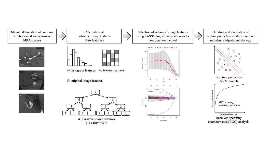
Prediction of Intracranial Aneurysm Rupture Risk Using Non-Invasive Radiomics Analysis Based on Follow-Up Magnetic Resonance Angiography Images: A Preliminary Study
Yamanouchi Masayuki, Arimura Hidetaka, Kodama Takumi, Urakami Akimasa
Applied Sciences, 2022, 12(17), 8615
https://www.mdpi.com/2076-3417/12/17/8615
This study This is the first preliminary study to develop prediction models
for aneurysm rupture risk using radiomics analysis based on follow-up magnetic
resonance angiography (MRA) images. We selected 103 follow-up images from
18 unruptured aneurysm (UA) cases and 10 follow-up images from 10 ruptured
aneurysm (RA) cases to build the prediction models. This prediction model
with non-invasive MRA images could predict aneurysm rupture risk for SAH
prevention.
Keywords: intracranial aneurysms; rupture risk; prediction model; radiomics; magnetic
resonance angiography
Papers 2022-2-Kodama
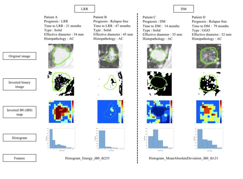
Relapse predictability of topological signature on pretreatment planning
CT images of stage I non-small cell lung cancer patients before treatment
with stereotactic ablative radiotherapy
Kodama Takumi, Arimura Hidetaka, Shirakawa Yumi, Ninomiya Kenta, Yoshitake
Tadamasa, Shioyama Yoshiyuki
Thoracic Cancer, 16 June, 2022
https://doi.org/10.1111/1759-7714.14483
This study aimed to explore the predictability of topological signatures
linked to the locoregional relapse (LRR) and distant metastasis (DM) on
pretreatment planning computed tomography images of stage I non-small cell
lung cancer (NSCLC) patients before treatment with stereotactic ablative
radiotherapy (SABR)
Keywords: lung cancer, topology, stereotactic ablative radiotherapy
Papers 2022-1-Ninomiya

Synergistic combination of a topologically invariant imaging signature
and a biomarker for the accurate prediction of symptomatic radiation pneumonitis
before stereotactic ablative radiotherapy for lung cancer: A retrospective
analysis
Kenta Ninomiya, Hidetaka Arimura, Tadamasa Yoshitake, Taka-aki Hirose, Yoshiyuki Shioyama
PLOS ONE, January 31, 2022
https://doi.org/10.1371/journal.pone.0263292
We aimed to explore the synergistic combination of a topologically invariant Betti number (BN)-based signature and a biomarker for the accurate prediction of symptomatic (grade ≥ 2) radiation-induced pneumonitis (RP+) before stereotactic ablative radiotherapy
(SABR) for lung cancer.
Keywords: Deep learning, Segmentation, Dense V-Networks, Lung stereotactic, Body
radiation therapy
a
Papers 2021-3-Urakami
Stratification of prostate cancer patients into low- and high-grade groups
using multiparametric magnetic resonance radiomics with dynamic contrast-enhanced
image joint histograms
Akimasa Urakami, Hidetaka Arimura, Yukihisa Takayama, Fumio Kinoshita,
Kenta Ninomiya, Kenjiro Imada, Sumiko Watanabe, Akihiro Nishie, Yoshinao
Oda, Kousei Ishigami
The Prostate, Published December /08/2021
DOI:https://doi.org/10.1002/pros.24278
This study aimed to investigate the potential of stratification of prostate
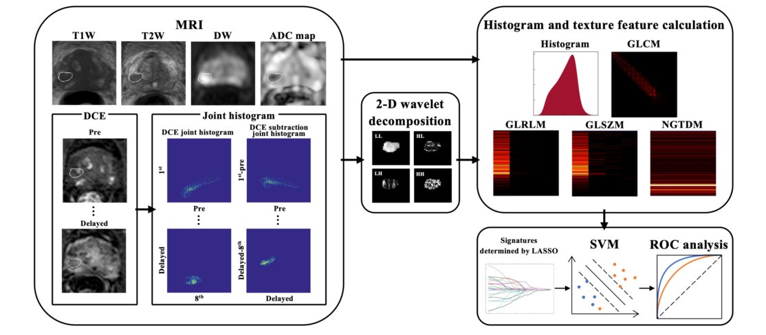
cancer patients into low- and high-grade groups (GGs) using multiparametric
magnetic resonance (mpMR) radiomics in conjunction with two-dimensional
(2D) joint histograms computed with dynamic contrast-enhanced (DCE) images.
This study suggests that the proposed approach could have the potential
to stratify prostate cancer patients into low- and high-GGs.
Keywords: prostate cancer, grade group, multiparametric MR, dynamic contrast-enhanced
images, joint histogram
PPapers 2021-2-Cui
Automated approach for segmenting gross tumor volumes for lung cancer stereotactic body radiation therapy using CT-based dense V-networks
Cui YunHao, Hidetaka Arimura, Risa Nakano, Tadamasa Yoshitake, Yoshiyuki
Shioyama, Hidetake Yabuuchi
Journal of Radiation Research, Volume 62, Issue 2, March 2021, Pages 346-355
Published : 22 January 2021
DOI:https://doi.org/10.1093/jrr/rraa132
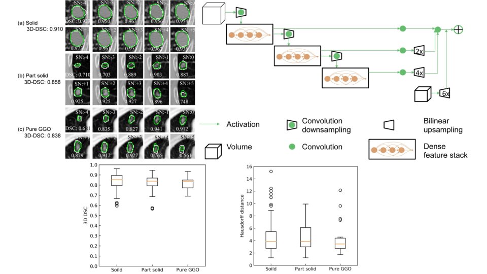
Abstract:The aim of this study was to develop an automated segmentation
approach for small gross tumor volumes (GTVs) in 3D planning CT images
using dense V-networks (DVNs) that offer more advantages in segmenting
smaller structures than conventional V-networks. Regions of interest (ROI)
with dimensions of 50 × 50 × 6-72 pixels in the planning CT images were
cropped based on the GTV centroids when applying stereotactic body radiotherapy
(SBRT) to patients.
Keywords: Deep learning, Segmentation, Dense V-Networks, Lung stereotactic, Body
radiation therapy
HPapers 2021-1-Ninomiya

Robust identification of EGFR mutated NSCLC patients from three countries using Betti numbers
Kenta Ninomiya, Hidetaka Arimura, Wai Yee Chan, Kentaro Tanaka, Shinichi Mizuno, Nadia Fareeda Muhammad Gowdh, Nur Adura Yaakup, Chong-Kin Liam, Chee-Shee Chai, Kwan Hoong Ng
Published by PLOS ONE, 11 January 2021
DOI:https://doi.org/10.1371/journal.pone.0244354
Abstract:We have proposed a novel robust radiogenomics approach to the
identification of epidermal growth factor receptor (EGFR) mutations among
patients with non-small cell lung cancer (NSCLC) using Betti numbers (BNs).
The proposed model showed higher robustness than the conventional models in the identification of EGFR mutations among NSCLC patients.
The results suggested the robustness of the BN-based approach against
Keywords:Homology, Radiogenomics, EGFR driver oncogene, Molecularly, Targeted therapy,
Imaging biopsy
Papers 2020-8-Le
Radiomic features based on Hessian index for prediction of prognosis in head-and-neck cancer patients
Quoc Le, Hidetaka Arimura, Kenta Ninomiya, Yutaro Kabata
Scientific Reports 10, Article number: 21301 (2020)
Published: 04 December 2020
DOI: https://www.nature.com/articles/s41598-020-78338-7
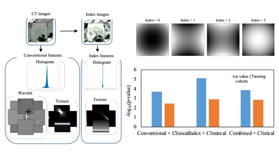
Purpose:This study proposed novel radiomic features based on the Hessian
index
of differential topology for the prediction of prognosis prior to treatment in head-and-neck (HN) cancer patients. The Hessian index, which can indicate tumor heterogeneity with convex, concave, and other points (saddle points), was calculated as the number of negative eigenvalues of the Hessian matrix at each voxel on computed tomography(CT)images.
Result:This result indicates that index features could provide more prognostic information than conventional features and further increase the prognostic value of clinical variables in HN cancer patients
Keywords:Head-and-neck cancer, Novel radiomics, CT images, Hessian index, Survival
analysis
Papers 2020-7-HiroseH
Radiomic prediction of radiation pneumonitis on pretreatment planning computed
tomography images prior to lung cancer stereotactic body radiation therapy
Taka-aki Hirose, Hidetaka Arimura, Kenta Ninomiya, Tadamasa Yoshitake,
Jun-ichi Fukunaga, Yoshiyuki Shioyama
Scientific Reports, 10, Article number: 20424(2020)
Published :24 November 2020
DOI:https://doi.org/10.1038/s41598-020-77552-7
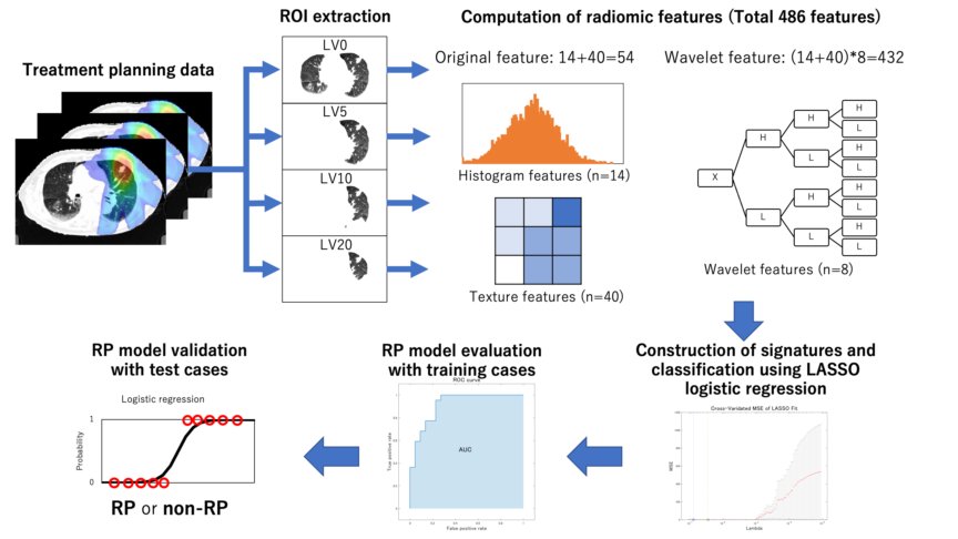
Abstract:This study developed a radiomics-based predictive model for radiation-induced pneumonitis (RP) after lung cancer stereotactic body radiation therapy (SBRT) on pretreatment planning computed tomography (CT) images. The radiomic features calculated on pretreatment planning CT images could be predictive imaging biomarkers for RP after lung cancer SBRT
Keywords:Radiomics-based predictive model, Radiation-induced pneumonitis, Lung
cancer stereotactic bodyradiation therapy, Pretreatment planning CT images,
Imaging biomarkers
Papers 2020-6-FitriaH
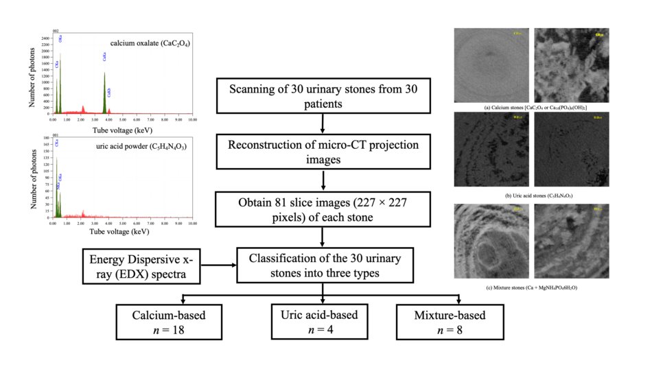
Automated classification of urinary stones based on microcomputed tomography images using convolutional neural network
Leni Aziyus Fitria, Freddy Haryanto, Hidetaka Arimura, Cui YunHao,Kenta
Ninomiya, Risa Nakano, Mohammad Haekal, Yuni Warty, Umar Fauzi
Physica Medica: European Journal of Medical Physics, Volume 78 Page 201-208
Published:08 October 2020
DOI: https://doi.org/10.1016/j.ejmp.2020.09.007
Purpose:The classification of urinary stones is important prior to treatment
because the treatments depend on three types of urinary stones, i.e., calcium,
uric acid, and mixture stones. We have developed an automatic approach
for the classification of urinary stones into the three types based on
microcomputed tomography (micro-CT) images using a convolutional neural
network (CNN).
Conclusion:The proposed automated CNN-based approach could successfully classify urinary stones into three types, namely calcium, uric acid, and mixture stones, using micro-CT images.
Keywords:Convolutional neural networkEnergy dispersive X-ray spectraMicro-CTUrinary
stones
Papers 2020-5-HossainH
Automated Approach for Estimation of Grade Groups for Prostate Cancer based
on Histological Image Feature Analysis
Alamgir Hossain, Hidetaka ARIMURA, Fumio Kinoshita, Kenta Ninomiya, Sumiko Watanabe, Kenjiro Imada, Ryoma Koyanagi, Yoshinao Oda
The Prostate, Volume 80 Issue 3 Page 291-302,
Published: 15 February 2020
DOI:10.1002/pros.23943
Background: There is a low reproducibility of the Gleason scores that determine
the grade group of prostate cancer given the intra‐ and interobserver variability
among pathologists.
This study aimed to develop an automated approach for estimating prostate cancer grade groups based on features obtained from histological image analysis.
Conclusions: Our results suggest that the proposed approach may support pathologists during the evaluation of grade groups for prostate cancer, thus mitigating intra‐ and interobserver variability.
Keywords:Gleason score, grade group, histological image features, International
Society of Urological Pathology (ISUP), piecewise step function
Papers 2020-4-NinomiyaH
Homological radiomics analysis for prognostic prediction in lung cancer patients
Kenta NINOMIYA, Hidetaka ARIMURA
Physica Medica: European Journal of Medical Physics, Volume 69 Page 90-100,
Published: 01 Januray 2020
DOI: https://doi.org/10.1016/j.ejmp.2019.11.026
Purpose: This study explored a novel homological analysis method for prognostic prediction in lung cancer patients.
Conclusion: This study demonstrates the excellent potential of HFs for prognostic prediction in lung cancer patients.
Keywords: Homology, Topologically invariant, Betti number, Radiomics, Lung cancer,
Survival prediction, Cox proportional hazard model
Papers 2020-3-HiroseH
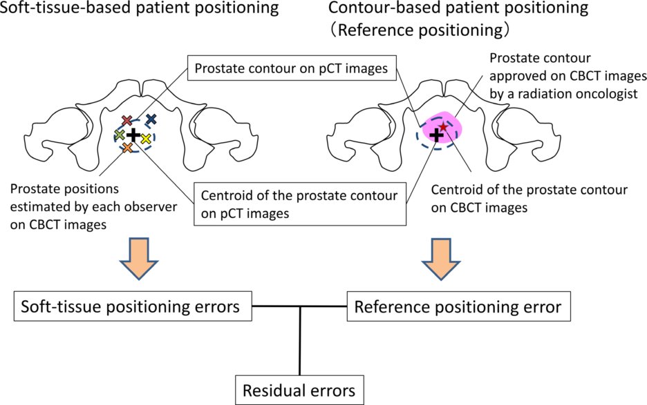
Observer Uncertainties of Soft Tissue-based Patient Positioning in IGRTwith artificial intelligence for precision medicine in radiation therapy
Taka-aki Hirose, Hidetaka Arimura, Jun-ichi Fukunaga, Saiji Ohga, Tadamasa
Yoshitake, Yoshiyuki Shioyama
Journal of Applied Clinical Medical Physics, Volume 21, Issue 2, Pages: 73-81, February 2020
Doi:org/10.1002/acm2.12817
Purpose: There remain uncertainties due to inter‐ and intraobserver variability in soft‐tissue‐based patient positioning even with the use of image‐guided radiation therapy (IGRT). This study aimed to reveal observer uncertainties of soft‐tissuebased patient positioning on cone‐beam computed tomography (CBCT) images for prostate cancer IGRT.
Conclusion: Intraobserver variability was sufficiently small and would be negligible. However, uncertainties due to interobserver variability for soft‐tissue‐based patient positioning using CBCT images should be considered in CTV‐to‐PTV margins.
Keywords:interobserver variation, intraobserver variation, prostate cancer image‐guided
radiation therapy, PTV margin, soft‐tissue‐based patient positioning.
Papers 2020-2-KaiH
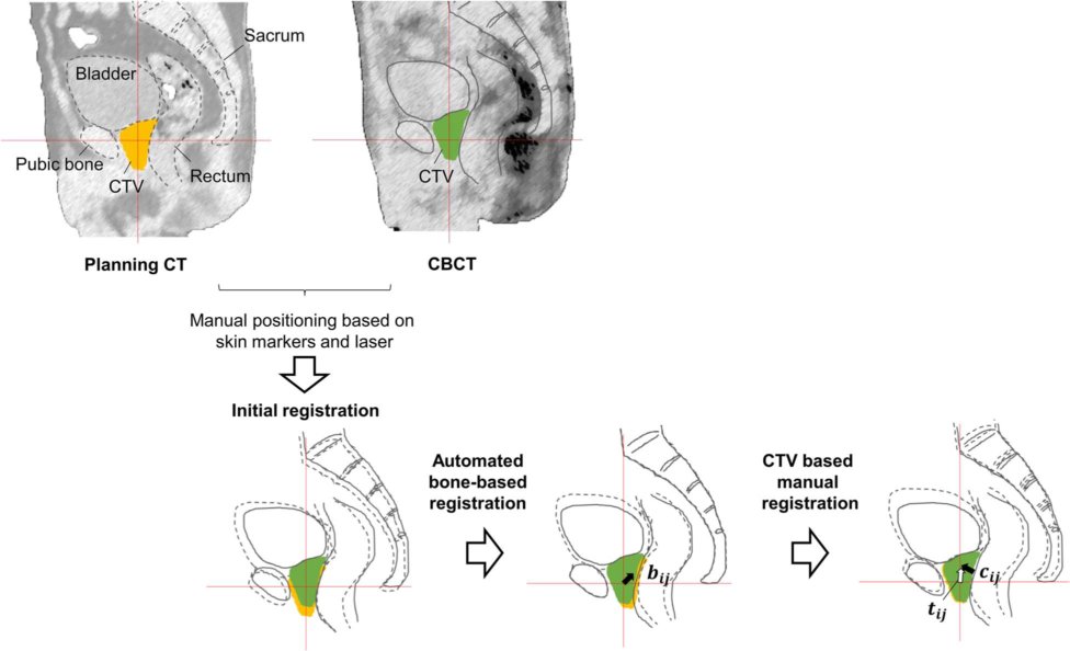
Semi-automated prediction approach of target shifts using machine learning
with anatomical features between planning and pretreatment CT images in
prostate radiotherapy
Yudai Kai, Hidetaka Arimura, Kenta Ninomiya, Tetsuo Saito, Yoshinobu Shimohigashi,
Akiko Kuraoka, Masato Maruyama, Ryo Toya, Natsuo Oya
Journal of Radiation Research,Volume 61, Issue 2, March 2020, Pages 285-297
Publisehd: 29 Januray 2020
Doi.org/10.1093/jrr/rrz105
The goal of this study was to develop a semi-automated prediction approach
of target shifts using machine learning architecture (MLA) with anatomical
features for prostate radiotherapy. Our hypothesis was that anatomical
features between planning computed tomography (pCT) and pretreatment cone-beam
computed tomography (CBCT) images could be used to predict the target,
i.e. clinical target volume (CTV) shifts, with small errors.
In conclusion, this study developed a semi-automated prediction approach
to CTV shifts using five types ofMLAs with anatomical features between
pCT and pretreatment CBCT images for improvement of the positioning PCa
patients in IGRT.
Papers 2020-1-Haseai

Similar-cases-based planning approaches with beam angle optimizations using water equivalent path length for lung stereotactic body radiation therapy
Shu Haseai, Hidetaka Arimura, Kaori Asai, Tadamasa Yoshitake, Yoshiyuki Shioyama
Radiological Physics and Technology, 13, 119-127,(2020)
Published: 14 March 2020
Doi.org/10.1007/s12194-020-00558-3
This study aimed to propose automated treatment planning approaches based
on similar cases with beam angle optimizations using water equivalent path
length (WEPL) to avoid lung and rib doses for lung stereotactic body radiation
therapy (SBRT)
This study indicates a potential of similar cases, whose beam angle configurations were optimized with WEPL to avoid lung and rib doses in lung SBRT plans.
Keywords Automated treatment planning, Similar cases, Lung stereotactic body radiation
therapy, Optimization, Water equivalent path length
Review paper
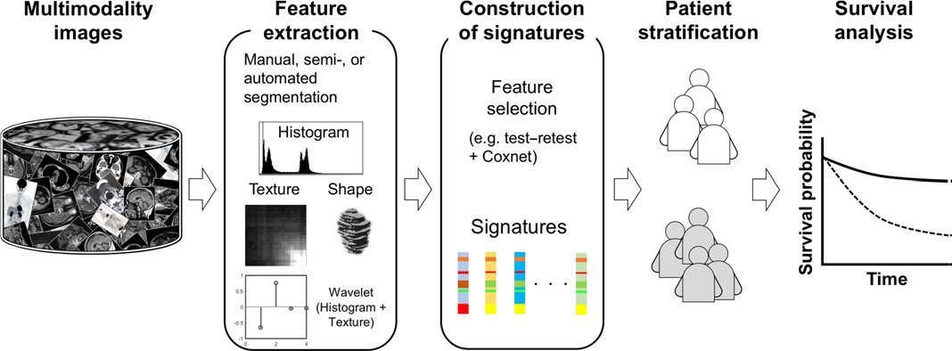
Radiomics with artificial intelligence for precision medicine in radiation therapy
Hidetaka Arimura, Mazen Soufi, Hidemi Kamezawa, Kenta Ninomiya,Masahiro
Yamada
Journal of Radiation Research, Vol. 60, Issue 1, January 2019, pp. 150-157, 2019.01
Publshed: 22 September 2018
Doi: 10.1093/jrr/rry077
Recently, the concept of radiomics has emerged from radiation oncology. It is a novel approach for solving the issues of precision medicine and how it can be performed, based on multimodality medical images that are noninvasive, fast and low in cost.
Radiomics is the comprehensive analysis of massive numbers of medical images in order to extract a large number of phenotypic features (radiomic biomarkers) reflecting cancer traits, and it explores the associations between the features and patients’ prognoses in order to improve decision-making in precision medicine.
Therefore, radiomic approaches, in combination with AI, may potentially enable practical use of precision medicine in radiation therapy by predicting outcomes and toxicity for individual patients.
Keywords: radiomics; artificial intelligence; precision medicine; radiation therapy;
medical images; cancer traits
Books
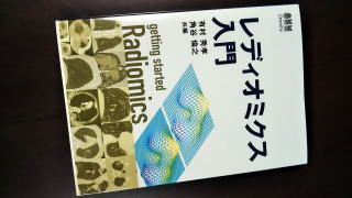
「レディオミクス入門」
著者:有村 秀孝 編、角谷 倫之 編
発売日:2021/10/19、発行元:オーム社
ISBN978-4-274-22638-0
医療分野にもAIが本格的に導入されつつある中で、AIを活用して網羅的な解析を行うレディオミクスが注目されています。本書は,レディオミクスについて系統的にまとめた初めての書籍です
レディオミクスの概要、レディオミクスの応用例、各応用例の精度、オープンソフトウェア紹介などで構成しています。レディオミクスは複雑な概念を含むので、図を多数掲載した、わかりやすいレディオミクスの入門テキストです。
H

「放射線治療AIと外科治療AI 医療AIとディープラーニングシリーズ」
著者:藤田広志シリーズ監修、有村秀孝編、諸岡健一編、2020/04/21、発行元:オーム社
分担執筆:「はじめに」、「Ⅱ放射線治療AI編 Chapter 1 放射線治療AIの概要」
ISBN978-4-274-22547-5
放射線治療と外科治療に関して、最新の内容をコンパクトにまとめてあります。
現在進行形のテーマをとりあげ現状と今後の進展について初学者にもわかるように、AI技術の必要性から始めてディープラーニングだけでなく、広くAI技術(機械学習を含む)を使った内容を中心に紹介しています。
A
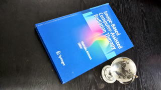
Image-Based Computer-Assisted Radiation Therapy
Edited by Hidetaka Arimura
Springer, April 2017
ISBN:978-981-10-2943-1
https://doi.org/10.1007/978-981-10-2945-5
This book provides a comprehensive overview of the state-of-the-art computational intelligence research and technologies in computer-assisted radiation therapy based on image engineering. It also traces major technical advancements and research findings in the field of image-based computer-assisted radiation therapy.
In high-precision radiation therapies, novel approaches in image engineering including computer graphics, image processing, pattern recognition, and computational anatomy play important roles in improving the accuracy of radiation therapy and assisting decision making by radiation oncology professionals, such as radiation oncologists, radiation technologists, and medical physicists, in each phase of radiation therapy.
All the topics presented in this book broaden understanding of the modern
medical technologies and systems for image-based computer-assisted radiation
therapy. Therefore this volume will greatly benefit not only radiation
oncologists and radiologists but also radiation technologists, professors
in medical physics or engineering, and engineers involved in the development
of products to utilize this advanced therapy.
Others
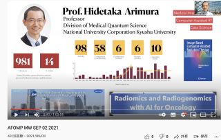
AFOMP Monthly Webinar, Sept 2, 2021: Radiomics and Radiogenomics with AI for Oncology
Topi: Radiomics and Radiogenomics with AI for Oncology
Speaker: Dr Arimura Hidetaka, Moderator: Dr. Hui-Yu Tsai
Hide Arimura Lab
Hidetaka Arimura, PhD
Division of Medical Quantum Science, Department of Health Sciences, Faculty of Medical Sciences,
Kyushu University
3-1-1, Maidashi, Higashi-ku, Fukuoka 812-8582, Japan